Digital radiography replaces traditional film with electronic sensors and computer processing to capture dental images. Instead of waiting for film to develop, the sensor records X-ray data and transfers it directly to a computer where a clear image appears within seconds. This shift from analog to digital streamlines diagnostics and makes imaging a routine, integrated part of modern dental care.
Sensors come in several styles — intraoral plates for bitewing and periapical views, and extraoral panels for broader imaging needs. Regardless of the device, the end result is the same: a high-resolution image that can be enhanced, measured, and archived without physical film or chemicals. For patients, this technology means faster appointments and more collaborative treatment planning between clinicians.
Because images are created and stored digitally, they become part of each patient’s electronic record. That allows clinicians to compare past images side-by-side with current ones, track changes over time, and document treatment outcomes. These capabilities help clinicians make more informed recommendations and tailor care to the individual patient.
One of the most significant advantages of digital radiography is reduced radiation exposure. Digital sensors are more sensitive than film, so they require less X-ray energy to produce diagnostically useful images. Dental teams follow safety protocols that limit radiation to the lowest level necessary while still obtaining clear diagnostic information.
Radiation safety is a standard part of the imaging process: protective measures such as lead aprons, thyroid collars when appropriate, and well-maintained equipment are used consistently. Clinicians also apply the ALARA principle — “as low as reasonably achievable” — to ensure patients receive only the imaging necessary for planning care and monitoring oral health.
For patients who are pregnant, very young, or have special health considerations, staff will discuss the need for radiographs and take extra precautions. When imaging is essential for diagnosis or treatment, digital radiography helps clinicians balance the need for information with a commitment to patient safety.
Digital images appear instantly on-screen, giving clinicians immediate visual feedback during an exam. This speed allows for on-the-spot adjustments when needed — for example, retaking an image at a different angle — without subjecting a patient to prolonged appointment time. Instant viewing also supports same-day decision-making for many common diagnostic needs.
Beyond speed, modern dental software offers tools to enhance diagnostic detail. Clinicians can adjust brightness and contrast, zoom in to inspect fine structures, and apply filters that reveal subtle differences in tissue density. These tools make it easier to detect early decay, evaluate root anatomy for endodontic care, and assess bone levels for periodontal and implant planning.
Because images can be annotated and measured precisely, they support accurate treatment planning. Measurements taken from digital radiographs help clinicians determine lesion size, root length, or the dimensions needed for restorative work — all without resorting to guesswork.
Digital radiography simplifies sharing images with other members of a patient’s care team. When a referral to a specialist is needed, high-quality digital images can be transmitted quickly and securely, helping referral appointments start from a well-informed place. Simultaneous access to images also allows multiple clinicians to consult on complex cases without delay.
Storing images electronically reduces physical clutter and the environmental impact associated with film processing. Digital archiving makes it easy to retrieve previous images for comparison or to include imaging documentation in treatment summaries. These practices support continuity of care and improve long-term record accuracy.
Maintaining the privacy and integrity of digital records is a priority. Practice systems are configured to protect patient data through secure storage, controlled access, and routine backups. These safeguards ensure that imaging data remains available to authorized clinicians while complying with applicable privacy standards.
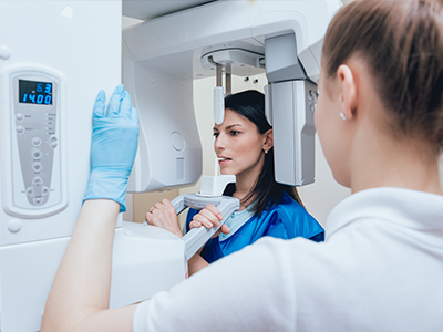
For most patients, a digital radiography appointment is quick and comfortable. A small sensor is positioned inside the mouth for intraoral views, or an external panel is placed for broader images. The clinician will explain each step and help position the sensor to minimize discomfort. Exposure itself lasts only a fraction of a second, and the team typically reviews the resulting image with the patient right away.
Special considerations are made for children and patients with gag reflex sensitivity or physical limitations. Clinicians use pediatric-sized sensors when appropriate and employ positioning techniques that keep imaging efficient and as comfortable as possible. Communication and patient comfort are integral parts of the imaging workflow.
The accuracy of digital imaging depends on both technology and people. Regular calibration of sensors, adherence to imaging protocols, and continuing education for the dental team help maintain high diagnostic standards. Our office routinely updates equipment and trains staff so images remain reliable tools for diagnosis and treatment planning.
At Dental Excellence of Brandon, we view digital radiography as one element of comprehensive, patient-centered care. If you have questions about why a particular image is recommended or what the results mean for your treatment, please contact us to learn more. We’re happy to explain our approach and how imaging supports safe, effective dental care.